

Protect Your Eyes With Regular Eye Exams
Undiagnosed and untreated eye diseases can lead to vision impairment and eventual blindness. There are about 1 in 28 Americans over age 40 living with low vision or blindness. Indeed, the leading causes of blindness and low vision in the United States are age-related eye diseases like age-related macular degeneration, cataracts, glaucoma, and diabetic retinopathy.
Each of these diseases is detectable during routine, comprehensive eye exams. And, although serious, early detection and early treatment can prevent potential vision loss.
If it has been more than a year since your last exam, or if you have been diagnosed with an ocular condition and need assistance managing it, please book an appointment with us today.
Why Early Detection Matters for Eye Disease
Many eye diseases develop without noticeable symptoms during their early stages. This means that an undetected eye disease could be doing permanent damage to your eye health without you realizing it.
In many cases, the damage caused by eye diseases, such as age-related macular degeneration or glaucoma, cannot be reversed. However, when detected early and managed carefully, we may be able to preserve your vision and prevent or delay vision loss from occurring.
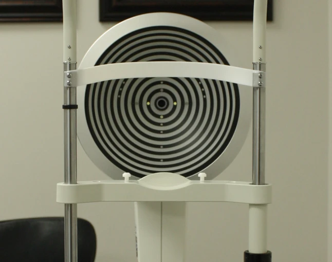
During an eye exam, we use our diagnostic technology to get a detailed picture of your eye health. These images can help us detect early signs of eye disease before you experience symptoms.
What Is Age-Related Macular Degeneration (AMD)?
While macular degeneration can happen at any age, it is the leading cause of severe vision loss in individuals over the age of 50. True to its namesake, age is a major risk factor for AMD.
AMD is a progressive deterioration of the macula, which is the part of your retina responsible for your central vision. As the macula wears out (due to age), hazy, fuzzy, or opaque spots form in your central vision.
AMD symptoms include:
- Distortion of straight lines, especially near central vision
- Fuzzy, dark, or white spots appear in central vision
- Worse or different color vision (rare)
During a comprehensive eye exam, we search for signs of AMD using optical coherence tomography (OCT). This extremely precise imaging method measures your eyes’ structures and cells to within 5 microns of accuracy. In conjunction with OCT, we use an extremely detailed retinal camera to take high-resolution images of the inside of your eye.
With these technologies, we get a clear depiction of your eye and the stage of your AMD.
There are two types of age-related macular degeneration: dry and wet.
- Dry AMD accounts for approximately 90% of cases of AMD. As the macular cells begin to age, their properties change, and they begin to cease functioning. Drusen, yellow deposits of fatty proteins, form underneath the retina and impair vision.
- Wet AMD is more severe than dry AMD. It occurs when weak blood vessels that form under the retina begin to leak blood and other fluids into the macula. This has a serious effect on vision and can be quite damaging, causing permanent vision loss.
What Are Cataracts?
Nearly 90% of Americans have developed a cataract by age 65, and the probability increases the older we get. An estimated 20.5 million Americans age 40 and older have cataracts in one or both eyes.
Cataracts are the clouding of the eyes’ natural lens, which leads to impaired vision and potential blindness. While they are commonly related to the aging process, there are different kinds of cataracts that can affect people of any age.
Cataract symptoms include:
- Blurry or cloudy vision, like looking through lightly-frosted glass
- Halos and glare from lights, especially when driving at night
- Changes in your color perception
- Frequent changes in your eyeglass or contact lens prescription
Cataracts progress slowly and are manageable in their early stages with updated prescription eyeglasses or contact lenses. They will continually worsen over time. When they start to impact your ability to read, write, drive, or perform other daily tasks, you may qualify for cataract surgery.
We will diagnose your cataracts during a comprehensive eye exam. This exam uses dilation, so be sure to arrange for transportation home.
What Is Glaucoma?
Glaucoma refers to a group of conditions that lead to the progressive damage of the optic nerve, which is responsible for transmitting information from the eyes to the brain. There are many different types and causes of glaucoma. Glaucoma is commonly related to increased internal eye pressure, but not always.
Glaucoma has earned the nickname of “the silent thief of sight” because it causes severe and irreversible eye damage before any noticeable vision loss. Glaucoma often erodes vision gradually, starting with side (peripheral) vision, so it may be difficult to notice it until significant vision loss has occurred.
An essential part of managing glaucoma is understanding its depth of progression. Our diagnostic technologies allow our eye care team to take precise, high-definition photos of your inner eye and optic nerve.
This intimate understanding of your eye health allows Dr. Baldwin and Dr. Nola to create and carefully monitor your personalized treatment plan.
Most forms of glaucoma do not have noticeable symptoms until significant peripheral vision is lost.
One version of glaucoma—acute angle-closure glaucoma—presents serious symptoms and is a medical emergency. Symptoms include:
- Severe headache
- Hazy, blurred vision
- Sudden eye pain
- Halos around bright lights
- Nausea or vomiting
- Sudden loss of sight in one or both eyes
Other Common Conditions We Can Diagnose and Manage
Beyond the eye diseases above, we also monitor and manage other eye conditions, ranging from acute eye infections to chronic conditions. While we monitor for most of these conditions through regular eye exams, if you notice a significant change in your vision, please book an appointment with us right away.
Conjunctivitis, also commonly known as pink eye, is the inflammation or infection of the conjunctiva, the clear tissue covering the whites of the eyes and inner eyelids.
Pink eye can be uncomfortable, irritating, and painful, but it rarely affects vision and likely won’t lead to long-term consequences. It can be caused by a bacterial or viral infection, which is very contagious, or as a result of an allergic reaction or chemical exposure.
Early diagnosis and treatment can help reduce the severity of your symptoms, alleviate discomfort, and address the condition accurately, while minimizing the spread of contagious conjunctivitis.
Dry eye occurs when your eyes don’t produce enough tears, or the tears evaporate too quickly. It can cause uncomfortable symptoms like burning, grittiness, redness, and intermittent blurred vision. In addition to being a distraction from daily life, dry eye can also lead to damage to the surface of your eye from improper lubrication.
We evaluate the quality and quantity of your tear film to address symptoms, so your eyes stay properly lubricated and healthy.
Floaters are the squiggly lines that float in our vision, while flashes are the spots or streaks of light that sometimes appear in our vision. These two visual phenomena are common and generally not medically dangerous.
However, the sudden onset of flashes or floaters may be a sign of a more serious condition. Always mention your flashes and floaters during your eye exams, just so your optometrist can rule out harmful eye conditions. And book an appointment right away if you notice any significant, sudden changes in your vision.
Keratoconus is an eye condition that causes the normally round cornea (the clear dome in front of your pupil and iris) to thin and bulge outward into a cone shape. This change can distort your vision.
Keratoconus is hereditary and more common in individuals with asthma, allergies, Ehlers-Danlos syndrome, Down syndrome, or retinitis pigmentosa. Symptoms include slight blurring and distortion of vision and light sensitivity, which typically develop in the late teen years or early 20s.
We can fit patients with keratoconus with special eyewear options to correct their vision, including scleral contact lenses, which have a unique, large-diameter design for a more comfortable fit. We may also refer out for corneal crosslinking, as necessary.
Visit Us in Wichita for the Management and Treatment of Eye Conditions
Managing your eye health doesn’t have to feel overwhelming. At The Eye Gallery, we combine a compassionate, patient-first approach with our noninvasive technology to safeguard your vision. Our diagnostic tools provide quick, painless scans to catch subtle changes early, helping prevent eye diseases and manage existing conditions without the stress.
Routine preventive care is your first line of defense, and our team is here to put you at ease. Let’s work together to preserve your sight.

What We Do
Our Services




Our Featured Eyewear Lines









Visit Our Location in Wichita
Our Address
Contact Information
- Phone: 316-315-0321
- Email: info@eyegalleryks.com
Hours of Operation

Kind Words
Patient Testimonials
test
Test
test
test
test
test
Our Google Reviews